-85%
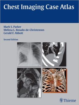
Exquisite Illustrations and Extensive Case Studies Define “Chest Imaging Case Atlas, Second Edition”
“Chest Imaging Case Atlas, Second Edition” stands out as an invaluable diagnostic resource for radiology residents, fellows, and practicing physicians, meticulously curated by renowned experts in chest imaging. This comprehensive atlas empowers readers to refine their diagnostic prowess by providing an unparalleled collection of over 200 case studies spanning a vast spectrum of conditions.
Illustrative Masterpiece:
The atlas is a visual feast, boasting over 1500 high-quality radiographs, 64-MDCT CT scans, multiplanar CT, CT angiographic visuals, and a selection of MRI and 3-D images. These exceptional images, captured with meticulous precision, provide an immersive visual experience that brings anatomical structures and pathological findings to life.
In-Depth Case Analyses:
Each case is meticulously dissected, presenting a thorough discussion of the disease in question, its underlying pathology, and its characteristic imaging manifestations. Typical and atypical findings are illustrated, providing a comprehensive understanding of each disorder. Additionally, the atlas offers valuable insights into management and prognosis, ensuring a multifaceted grasp of the clinical implications.
Cutting-Edge Advancements:
This updated edition incorporates the latest advancements in imaging technology, seamlessly integrating more multiplanar, CT angiographic (CTA), and MR images throughout the text. This ensures that readers remain at the forefront of this rapidly evolving field. A testament to its currency, the atlas proudly boasts 40 fresh cases, bolstering its comprehensive coverage.
Post-Thoracic Interventions:
A novel section dedicated to post-thoracotomy chest imaging equips readers with the necessary knowledge to interpret normal post-operative findings and identify potential complications associated with various thoracic interventional procedures.
Cardiovascular Enhancements:
The adult cardiovascular disease section has been significantly expanded, addressing the complex challenges of univentricular and biventricular heart failure, including various ventricular assist devices and the Total Artificial Heart. The atlas provides a comprehensive overview of their imaging characteristics, enabling confident diagnosis and meticulous management.
Refined Pulmonary Insights:
The diffuse lung disease section has been meticulously expanded, incorporating an innovative approach to HRCT interpretation. This empowers readers to master the complexities of this advanced technique, unlocking a deeper understanding of pulmonary pathologies.
Sharpening Diagnostic Skills:
Throughout the atlas, insightful pearls pinpoint diagnostic features that provide compelling support for specific diagnoses. These invaluable nuggets of wisdom fine-tune clinical acumen, enabling readers to navigate diagnostic challenges with confidence.
Indispensable Reference:
“Chest Imaging Case Atlas, Second Edition” stands as an indispensable illustrated reference for anyone involved in radiology and chest imaging, including pulmonary medicine physicians, thoracic surgeons, and radiologists. Its unparalleled coverage, cutting-edge imagery, and insightful case analyses make it an essential resource for both residents and experienced practitioners alike.




Reviews
Clear filtersThere are no reviews yet.