-56%
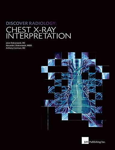
Understanding the Structure of a Chest X-Ray Textbook
Section I: The Fundamentals of Chest X-Ray Interpretation
This section delves into the foundational principles of chest X-ray analysis, providing a comprehensive understanding of the imaging modality and the process involved in deciphering it.
Chapter 1: The Essence of a Chest X-Ray
This chapter lays the groundwork by defining the purpose and significance of chest X-rays, explaining how they are acquired and the various factors that influence their appearance.
Chapter 2: The X-Ray Image: A Visual Portrait
This chapter examines the subtleties of X-ray images, exploring the concepts of density, contrast, and the interplay of different tissues and structures within the chest cavity.
Chapter 3: The Unveiling of the Interpretive Process
This chapter introduces the systematic approach to chest X-ray interpretation, highlighting the crucial steps involved in identifying and analyzing abnormalities.
Chapter 4: Imaging the Thoracic Framework
This chapter delves into the radiological anatomy of the thorax, providing a detailed understanding of the bones, muscles, and mediastinal structures visible on a chest X-ray.
Chapter 5: Recognizing Pathology’s Imprint
This chapter elucidates the concept of radiological zones, enabling readers to differentiate between normal and pathological findings in the chest.
Section II: Delving into Chest Anatomy through Radiological Lenses
This section transports readers into the intricate world of chest anatomy as seen through the lens of X-rays, connecting radiological observations to the underlying structures.
Chapter 6: The Pulmonary Panorama
This chapter embarks on an in-depth exploration of the lungs, examining their radiological appearance in both normal and pathological states.
Chapter 7: The Heart’s X-Ray Silhouette
This chapter focuses on the radiological anatomy of the heart, describing its various chambers, valves, and vessels as they appear on chest X-rays.
Chapter 8: The Pleural Envelope and Beyond
This chapter delves into the anatomy of the pleura, mediastinum, and diaphragm, highlighting their significance in chest X-ray interpretation.
Chapter 9: The Radiological Symphony of Chest Structures
This chapter culminates the section by weaving together the radiological anatomy of individual structures, demonstrating how they combine to form the comprehensive image of the thorax.
Section III: Applying the Interpretative Toolkit
This section empowers readers to apply their newfound knowledge and skills to real-world chest X-ray interpretation, guiding them through the process of identifying and localizing pathology with confidence.
Chapter 10: Systematic Scanning for Pathological Clues
This chapter imparts a structured approach to chest X-ray analysis, ensuring thorough and accurate evaluation of all pertinent areas.
Chapter 11: Localizing Pathology: Precision and Accuracy
This chapter sharpens readers’ ability to pinpoint the exact location of abnormalities on chest X-rays, employing a combination of techniques and anatomical landmarks.
Chapter 12: Embracing Digital Technologies in Interpretation
This chapter explores the latest digital technologies available in chest X-ray interpretation, discussing their capabilities and limitations.
Chapter 13: When to Request a Chest X-Ray: Clinical Decision-Making
This concluding chapter provides practical guidance on when a chest X-ray is an appropriate diagnostic tool, considering clinical symptoms and other relevant factors.
maybe you like these too:
- Chest X-Rays for Medical Students: CXRs Made Easy, 2nd Edition (Original PDF from Publisher)
- Radiopedia 2021 (July 19 – 23) (Videos, Well Organized)
- Chest Radiographic Interpretation in Pediatric Cardiac Patients (ORIGINAL PDF from publisher)
- Imaging of the Lower Extremity, An Issue of Radiologic Clinics of North America The Clinics: Radiology) (Original PDF from Publisher)



Reviews
Clear filtersThere are no reviews yet.