-68%
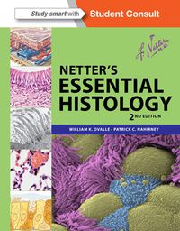
Netter’s Essential Histology: A Comprehensive Guide to Microscopic Anatomy
Netter’s Essential Histology provides an unparalleled synthesis of gross anatomy, embryology, and cutting-edge microscopic imaging techniques to empower students and practitioners alike with an in-depth understanding of histological principles and their implications for clinical practice.
Visual Masterpieces
This comprehensive histology textbook-atlas features a remarkable collection of visual elements, including Netter’s iconic medical illustrations and a vast array of light and electron micrographs, carefully curated to unveil the intricacies of human tissues at the microscopic level. These captivating images serve as invaluable tools for mastering histological concepts and their clinical relevance.
Anatomical Foundation
Rooted in a strong anatomical framework, Netter’s Essential Histology ensures a seamless integration of microscopic observations with the macroscopic anatomy of the human body. This holistic approach enables readers to grasp the functional significance of histological structures and their role in maintaining homeostasis and responding to pathological processes.
Succinct Explanations
Throughout the textbook, concise explanatory text complements the visual presentations, providing clear and accessible insights into histological structures and processes. This user-friendly approach facilitates the assimilation of complex histological concepts, even for students encountering the subject for the first time.
Clinical Connections
Numerous clinical boxes strategically placed throughout the text highlight the practical relevance of histological findings in clinical settings. These insightful annotations provide a direct link between microscopic observations and real-world patient care, fostering a deeper understanding of the diagnostic and therapeutic implications of histological alterations.
Enhanced Electron Micrographs
In this latest edition, Netter’s Essential Histology introduces an expanded collection of electron micrographs, meticulously enhanced and colorized to reveal the ultra-fine details of cellular components in stunning three-dimensional detail. These state-of-the-art images provide an unprecedented glimpse into the molecular and subcellular processes that govern cellular function and behavior.
Interactive Learning Tools
To further enhance the learning experience, Netter’s Histology Flash Cards (sold separately) complement the textbook with over 200 concise, full-color cards featuring key histological concepts and corresponding images. These portable tools enable students to reinforce their understanding at their own pace, promoting active recall and efficient retention of histological knowledge.
maybe you like these too:
- 2017 Pathology Review Pulmonary, Neuro, and Hepatic Pathology for the General Pathologist (Videos)
- Brock Biology of Microorganisms, 14e (Original PDF from Publisher)
- Review of Medical Microbiology and Immunology, 13th Edition (EPUB)
- e-Study Guide for: Introduction to Pharmacology by Mary Kaye Asperheim Favaro, ISBN 9781416001898 (MOBI)


Reviews
Clear filtersThere are no reviews yet.