-33%
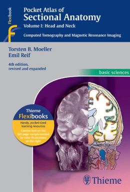
Pocket Atlas of Sectional Anatomy: Enhanced Edition for Precise Radiologic Interpretation
A Comprehensive and Invaluable Guide for Clinicians
[by Torsten Bert Moeller]**
With its unparalleled illustrations and meticulous depiction of sectional anatomy, this esteemed atlas serves as an indispensable navigational tool for clinicians seeking to master radiologic anatomy and proficiently interpret CT and MR images. This updated edition seamlessly integrates with its companion volumes to provide a highly specialized and comprehensive resource.
Key Features:
- Didactic Organization: Two-page units featuring high-quality radiographs juxtaposed with brilliant, full-color diagrams
- Extensive Imaging: Hundreds of meticulously rendered CT and MR images, captured with the latest advanced scanners
- Consistent Color-Coding: Facilitating the effortless identification of similar structures across multiple slices
- Concise Labeling: All figures are expertly labeled for clarity and ease of understanding
Expansions and Enhancements:
- Volume I Updates:
- Novel cranial CT imaging sequences of the axial and coronal temporal bone
- Expanded MR section, boasting new 3T MR images of the temporal lobe and hippocampus, basilar artery, cranial nerves, cavernous sinus, and more
- Additional arterial MR angiography sequences of the neck and larynx images
Benefits:
- Enhanced Visualization: Exceptional illustrations and images provide an immersive understanding of sectional anatomy
- Precision in Diagnosis: Accurate interpretation of CT and MR images leads to more informed clinical decision-making
- Versatility: Ideal for both clinical practice and study settings, offering quick recall and easy reference
The Pocket Atlas of Sectional Anatomy embodies a testament to the power of visual representation in medical imaging. Its meticulous attention to detail and comprehensive approach make it an invaluable asset for any clinician dedicated to excellence in radiologic interpretation.



Reviews
Clear filtersThere are no reviews yet.