-91%
The Immeasurable Value of Skeletal Maturation Atlases: An In-Depth Exploration
The significance of skeletal maturation atlases in the field of pediatrics cannot be overstated. These invaluable resources empower healthcare professionals with a comprehensive understanding of the intricate developmental milestones of the growing skeleton, guiding accurate interpretations and enhancing patient care.
Comprehensive Reference Points for Normal Skeletal Growth
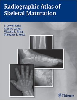
Radiographic Atlases of Skeletal Maturation offer an unparalleled collection of high-quality images that serve as benchmarks for normal skeletal development. These atlases meticulously catalog images of both male and female individuals across various age ranges and body regions. By providing multiple projections for each age, sex, and body part combination, these atlases facilitate direct comparisons between reference images and specific patient cases. This meticulous approach allows clinicians to pinpoint normal developmental expectations for any given pediatric skeletal radiograph.
Resolving Diagnostic Uncertainties with Confidence
The abundance of reference images in skeletal maturation atlases empowers clinicians to swiftly ascertain whether a particular image aligns with normal developmental patterns. This clarity is particularly crucial when evaluating pediatric skeletal radiographs in time-sensitive settings, such as emergency rooms or on-call consultations. For instance, when presented with a young patient suspected of an elbow injury, the atlas can assist in differentiating between a fracture and a normal ossification center, enabling an informed diagnosis.
Systematic Organization for Expedient Access
Skeletal maturation atlases are meticulously organized by gender and body part, ensuring rapid retrieval of age-specific developmental images. This user-friendly design allows clinicians to swiftly access the precise information they require, saving valuable time and facilitating accurate interpretations.
Advanced Image Manipulation Tools for Enhanced Clarity
Contemporary skeletal maturation atlases often incorporate sophisticated software packages equipped with image enhancement tools. These tools empower users to optimize atlas image details, enhancing clarity and facilitating precise evaluations. Compatibility with various image formats, including DICOM, allows for seamless integration with existing imaging systems and the viewing and editing of external images.
Invaluable Support for Pediatric Radiographic Interpretation
The comprehensive collection of images in skeletal maturation atlases proves indispensable for interpreting pediatric skeletal radiographs in any clinical setting. These atlases provide the full spectrum of comparative information needed to make confident diagnoses, becoming an indispensable resource for radiologists, pediatricians, orthopedists, emergency room physicians, internists, rehabilitation physicians, and trainees.
Conclusion
Skeletal maturation atlases have revolutionized the field of pediatric skeletal radiology, providing a comprehensive and accessible resource for healthcare professionals. Their ability to establish clear reference points for normal skeletal development and resolve diagnostic uncertainties with confidence makes them an indispensable tool for accurate and timely patient care.








Reviews
Clear filtersThere are no reviews yet.