-96%
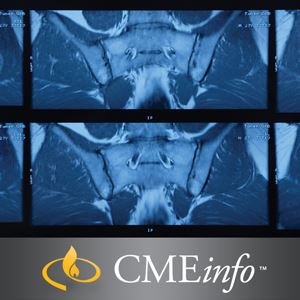
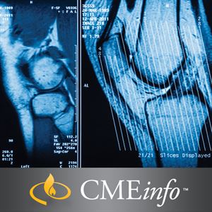
Musculoskeletal MRI: A Comprehensive Exploration for Advanced Imaging
This state-of-the-art musculoskeletal imaging program, led by the renowned University of California, San Francisco, presents a comprehensive overview of the intricate anatomy and pathological manifestations of various musculoskeletal structures.
Delving into the Musculoskeletal System
Participants will embark on an in-depth journey, exploring the complex anatomy and pathology of each joint, encompassing cartilaginous lesions, bone tumors, and more. This exploration will empower you with the knowledge needed to optimize your diagnostic skills in this specialized field.
Mastering Musculoskeletal Imaging Techniques
Through a comprehensive review of the pertinent anatomy and pathology of various joints, this program will provide you with the tools necessary to select the most appropriate imaging protocols. You will learn about the latest sequencing techniques for evaluating cartilage lesions, as well as optimized protocols utilizing both 1.5T and 3T MR units.
Beyond Joints: Exploring Bone and Soft Tissue Pathology
The program ventures beyond the confines of joints, delving into the realm of bone and soft-tissue tumors, as well as bone marrow and muscle pathology. This comprehensive approach will enhance your understanding of a wide range of conditions affecting the musculoskeletal system.
Unveiling Abnormalities on MRI
Participants will become adept at identifying abnormalities in joints, muscles, bone marrow, and cartilage on MRI, enabling them to assess their clinical significance. Additionally, they will gain the ability to recognize common bone tumors, developing a differential diagnosis approach to guide clinical management.
Cervical Spine Trauma: Understanding Complex Injuries
This program addresses the challenges of interpreting cervical spine trauma on MRI, enabling participants to identify suitable protocols and pinpoint abnormalities in this complex region.
Skill-Building Objectives
Upon completing this program, participants will be able to:
- Employ optimized imaging protocols for the musculoskeletal system, including suppression of metallic implants in post-arthroplasty patients
- Comprehend the intricate MRI anatomy of major joints, including the shoulder, digits, hip, knee, and ankle
- Detect and interpret MRI abnormalities in joints, muscles, bone marrow, and cartilage, understanding their clinical significance
- Recognize common bone tumors, developing a differential diagnosis
- Identify anomalies and potential pitfalls in cervical spine trauma imaging
Target Audience
This program is tailored for diagnostic radiologists, orthopedic surgeons, sports medicine physicians, and MRI technologists who possess a basic or intermediate understanding of musculoskeletal MRI and aim to elevate their knowledge to an advanced level.
Program Topics and Faculty
- ACL Reconstruction: Matthew D. Bucknor, MD
- Elbow Injuries in Athletes: Matthew D. Bucknor, MD
- Radiographic Checklist for Hip Impingement: Matthew D. Bucknor, MD
- Three Tough Topics in the Foot/Ankle: Matthew D. Bucknor, MD
- Digital MRI: The Fingers and Toes: John F. Feller, MD
- Imaging of Cervical Spine Trauma: John F. Feller, MD
- MRI Following Joint Arthroplasty: John F. Feller, MD
- Bone Marrow: Thomas M. Link, MD, PhD
- Bone Tumors: Thomas M. Link, MD, PhD
- Cartilage and Osteochondral Disease: Thomas M. Link, MD, PhD
- Inflammatory Arthropathies: Thomas M. Link, MD, PhD
- MRI of Pelvic Tendons: Mini N. Pathria, MD
- Nerve Entrapment: Mini N. Pathria, MD
- Stress Injury of Bone: Mini N. Pathria, MD
- Traumatic Muscle: Mini N. Pathria, MD
- Meniscal Pearls and Pitfalls: Lynne S. Steinbach, MD
- Pitfalls in Shoulder MRI: Lynne S. Steinbach, MD
- Traumatic Shoulder Instability: Lynne S. Steinbach, MD
Availability
Series Release Date: May 1, 2018
Series Expiration Date: April 30, 2021


Reviews
Clear filtersThere are no reviews yet.