-96%
Comprehensive Thoracic Imaging Review: Unveiling the Latest Advancements
Comprehensive Overview of Thoracic Imaging
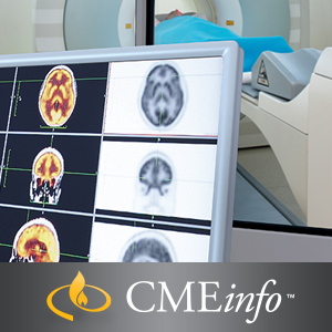
This CME unveils a comprehensive and multifaceted review of cross-sectional imaging of the chest, equipping attendees with the latest imaging methodologies and diagnostic techniques. UCSF Thoracic Imaging presents a deep dive into specialized areas, including cardiovascular imaging, pulmonary imaging, lung cancer screening, and practical applications of lung cancer staging.
Expert Insights into Thoracic Imaging
Cardiovascular Imaging:
- Unveiling cardiac findings on routine chest CT with Dr. Travis S. Henry, MD
- Imaging pulmonary hypertension: Advanced techniques and interpretation
- Introduction to coronary CTA: A comprehensive guide to its applications
- Acute aortic syndromes: Diagnostic algorithms and management strategies
Pulmonary Emboli:
– Understanding cross-sectional imaging in the accurate assessment of pulmonary embolism
– Perioperative aorta: Crucial imaging insights for surgical interventions with Dr. Michael D. Hope, MD
Lung Cancer and Other Tumors:
- Mediastinal masses: Differentiating benign from malignant lesions with Dr. Brett M. Elicker, MD
- Typical and atypical appearances of lung cancer: Enhancing diagnostic accuracy
- Practical applications of lung cancer staging: Optimizing treatment plans
- Incidentals and artifacts on PET: Avoiding pitfalls in interpretation
Pulmonary Imaging:
- Multi-disciplinary approach to diffuse lung disease: Unveiling underlying pathologies
- Hypersensitivity pneumonitis: Imaging pearls for accurate diagnosis
- Pulmonary infections: Key patterns for effective characterization
- Idiopathic pulmonary fibrosis: An update on diagnostic and management strategies
- HRCT findings: Comprehensive analysis of ground glass opacity, consolidation, mosaic perfusion, cysts, emphysema, nodular disease, reticular patterns, and fibrosis
Learning Objectives and Target Audience
Upon completion of this CME, participants will demonstrate proficiency in:
- Confidently interpreting high-resolution CT scans of the lungs, providing precise differential diagnoses
- Applying a practical approach to pulmonary infection imaging
- Distinguishing various aortic diseases based on imaging manifestations
- Utilizing cross-sectional imaging to effectively evaluate pulmonary embolism
- Differentiating benign and malignant lung lesions using CT and PET scans
- Understanding the crucial role of CT and PET in lung cancer evaluation and staging
This educational activity is specifically tailored for radiologists and other medical professionals seeking to enhance their expertise in thoracic image interpretation.




Reviews
Clear filtersThere are no reviews yet.