Understanding Thoracic Imaging: A Comprehensive Guide for Healthcare Professionals
This in-depth Continuing Medical Education (CME) program delves into the complexities of cross-sectional thoracic imaging, empowering healthcare providers with the latest clinical guidelines to enhance patient care.
Latest Advancements in Thoracic Imaging
Led by renowned thoracic imaging expert Travis S. Henry, MD, this program covers a comprehensive spectrum of topics, including:
Acute aortic syndromes
Pulmonary nodules
Lung cancer screening
Thoracic incidentalomas
Mosaic attenuation
Bronchiectasis imaging
By mastering these concepts, participants will gain invaluable insights into:
Interpreting high-resolution CT scans of the lungs for precise differential diagnoses
Utilizing a systematic approach to the imaging of lung infections
Identifying hallmark features of acute and chronic aortic diseases
Assessing the role of cross-sectional imaging in evaluating pulmonary embolism
Distinguishing benign from malignant lung nodules/masses via CT and PET
Expert Insights: Speakers and Topics
This CME program features a distinguished faculty of experts who provide invaluable insights into various aspects of thoracic imaging:
Cystic Lung Disease and Emphysema: Brett M. Elicker, MD
Mosaic Attenuation: Brett M. Elicker, MD
Multidisciplinary Approach to ILD: Brett M. Elicker, MD
Putting It All Together: Expert Reporting Techniques: Brett M. Elicker, MD
Update on UIP: Brett M. Elicker, MD
Imaging of Bronchiectasis: Travis S. Henry, MD
Imaging of the Pleura: Travis S. Henry, MD
Nodules and Small Airways Disease: Travis S. Henry, MD
Non-Thrombotic Pulmonary Emboli: Travis S. Henry, MD
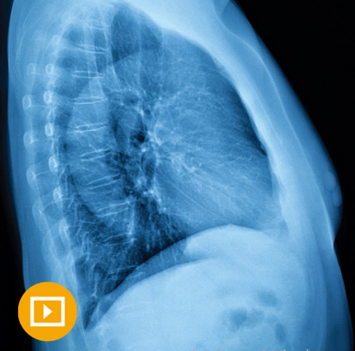
The Lateral Radiograph: Decoding Complexities: Travis S. Henry, MD
Acute Aortic Syndromes: Michael D. Hope, MD
Imaging of Pulmonary Embolism: Michael D. Hope, MD
The PA Radiograph: Mastering Interpretation: Michael D. Hope, MD
Cardiac Findings on Non-Gated CT: Kimberly G. Kallianos, MD
Mediastinal Masses: Kimberly G. Kallianos, MD
Post-operative Chest Imaging: Kimberly G. Kallianos, MD
Thoracic Incidentalomas: Kimberly G. Kallianos, MD
HRCT Basics: Ground-Glass and Consolidations: David M. Naeger, MD
Incidentals and Artifacts on PET/CT: Avoiding Misinterpretation: David M. Naeger, MD
Lung Cancer Screening: Adhering to Best Practices: David M. Naeger, MD
Pulmonary Nodules: Comprehensive Evaluation using CT and PET/CT: David M. Naeger, MD
Typical and Atypical Appearances of Lung Cancer: David M. Naeger, MD
Learning Objectives: Enhancing Diagnostic Skills
Upon completing this CME program, participants will be equipped with the knowledge and skills to:
Interpret high-resolution CT scans of the lungs, providing targeted differential diagnoses
Apply a practical approach to imaging lung infections
Recognize characteristic manifestations of acute and chronic aortic diseases
Leverage cross-sectional imaging for effective pulmonary embolism assessment
Distinguish benign from malignant lung nodules/masses using CT and PET
Utilize CT and PET in evaluating and staging lung cancer
This educational activity is tailored for radiologists and other healthcare professionals seeking to enhance their knowledge of thoracic image interpretation.
Program Availability: Access to Cutting-Edge Insights
The program was released on April 16, 2019, and will remain accessible until April 15, 2022.


Reviews
Clear filtersThere are no reviews yet.